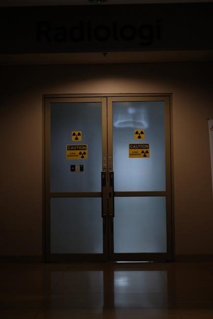
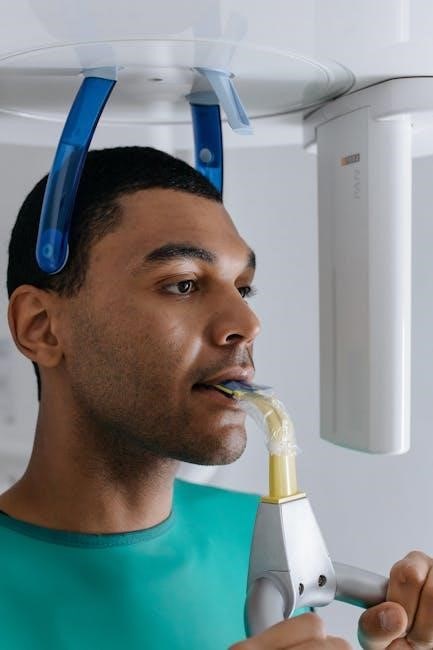
The 9th Edition of Radiation Protection in Medical Radiography provides foundational knowledge on radiation safety, emphasizing the balance between diagnostic quality and patient protection. It covers natural radiation exposure, regulatory guidelines, and advanced technologies to minimize risks in medical imaging, ensuring safe practices for both patients and healthcare workers.
1.1 Overview of the 9th Edition
The 9th Edition of Radiation Protection in Medical Radiography is a comprehensive guide covering 15 chapters. It addresses key topics such as types of radiation, biological effects, patient and personnel protection, and regulatory guidelines. Designed for radiologic technologists, the book simplifies complex concepts while maintaining depth. Authors Mary Alice Statkiewicz Sherer, Paula J. Visconti, E. Russell Ritenour, and Kelli Haynes ensure a practical approach to radiation safety. The edition emphasizes balancing diagnostic quality with patient protection, offering updated strategies for minimizing radiation exposure in medical imaging.
1.2 Importance of Radiation Protection in Medical Imaging
Radiation protection is vital to minimize risks for patients and healthcare workers. It ensures diagnostic quality while reducing exposure to ionizing radiation. Proper practices prevent long-term health effects like cancer and genetic mutations. The 9th Edition highlights the significance of balancing safety with image quality, emphasizing ethical considerations and regulatory compliance. By adhering to principles like ALARA, medical imaging can achieve optimal outcomes, safeguarding both patients and personnel from unnecessary radiation exposure.

Fundamentals of Radiation
Radiation is energy emitted as waves or particles. Ionizing radiation alters matter, while non-ionizing radiation, like light, does not. Both types interact with living tissues differently.
2.1 Types of Radiation: Ionizing and Non-Ionizing
Radiation is classified into ionizing and non-ionizing types. Ionizing radiation, such as X-rays and gamma rays, has high energy that can ionize atoms, potentially causing DNA damage. Non-ionizing radiation, like ultraviolet (UV) and radio waves, has lower energy and does not ionize atoms but can still cause thermal effects. Understanding these types is crucial for assessing risks and implementing safety measures in medical imaging. Both types interact with biological tissues differently, necessitating specific protective strategies to minimize exposure and ensure patient safety.
2.2 Sources of Radiation: Natural and Man-Made
Natural radiation sources include cosmic rays, radon gas, and radioactive elements in soil and water. Man-made sources involve medical imaging, nuclear power, and consumer products. Medical radiography uses X-rays, while natural background radiation, like cosmic rays, contributes to human exposure. Understanding these sources helps assess risks and implement safety measures. Natural radiation accounts for most annual exposure, while medical imaging is a controlled, essential man-made source. Balancing both is key to effective radiation protection strategies in healthcare and daily life.
Biological Effects of Radiation
Radiation exposure can cause acute effects like radiation sickness and long-term risks such as cancer and genetic mutations, depending on dose and duration of exposure.
3.1 Acute Radiation Syndrome (ARS)
Acute Radiation Syndrome (ARS) occurs after exposure to high doses of ionizing radiation, causing severe damage to the bone marrow, lungs, gastrointestinal system, and central nervous system. Symptoms include nausea, vomiting, diarrhea, fatigue, and even seizures or coma in extreme cases.
The onset of symptoms varies depending on the radiation dose, with higher doses leading to more rapid and severe effects. Recovery can take years, and lower doses may result in long-term health issues like cataracts and increased cancer risk. Understanding ARS is crucial for medical professionals to manage and prevent radiation-related illnesses effectively.
3.2 Long-Term Effects: Cancer and Genetic Mutations
Long-term radiation exposure can lead to cancer and genetic mutations, even at low doses. Studies show radiation-induced cataracts and increased cancer risks, with effects appearing years after exposure. Understanding these risks is vital for minimizing radiation exposure in medical settings, ensuring patient and worker safety through proper protection measures and adherence to safety guidelines.
Radiation Exposure in Medical Settings
Radiation exposure in medical settings is a critical concern, involving both natural and man-made sources. Minimizing radiation exposure is essential for patient and staff safety.
4.1 Patient Exposure: Dose Limits and Risk Assessment
Patient exposure to radiation in medical settings requires careful management to ensure doses remain within safe limits. Natural sources, such as cosmic rays, contribute significantly to annual exposure, averaging 2.4 mSv. Risk assessment involves evaluating the diagnostic benefits against potential harm, emphasizing justification and optimization. Dose limits are established to minimize long-term effects like cancer and genetic mutations. Strategies to reduce exposure include using lead aprons, thyroid collars, and modern imaging technologies that lower radiation doses while maintaining image quality.
4.2 Occupational Exposure: Radiation Safety for Healthcare Workers
Healthcare workers in radiography are exposed to radiation from various sources, including natural background radiation and man-made sources like X-rays. Annual doses for workers typically remain below regulatory limits, but consistent monitoring is crucial. The ALARA principle guides practices to minimize exposure, emphasizing time management, distance, and shielding. Personal protective equipment, such as lead aprons, and regular dosimetry monitoring ensure safety. Training and awareness programs further mitigate risks, ensuring adherence to safety protocols and fostering a culture of radiation protection in medical imaging environments.
Principles of Radiation Protection
Radiation protection balances patient and staff safety with image quality, ensuring minimal exposure while maintaining diagnostic accuracy. Guidelines emphasize optimization, dose limits, and continuous monitoring to prevent harm.
5.1 Time, Distance, and Shielding: The 3 Main Principles
The three main principles of radiation protection are time, distance, and shielding. Minimizing exposure time reduces radiation dose. Increasing distance from the source leverages the inverse square law, where radiation intensity decreases with distance. Shielding involves using materials like lead to block or absorb radiation. These principles, combined with the ALARA (As Low As Reasonably Achievable) concept, ensure radiation exposure is minimized while maintaining diagnostic image quality and patient safety in medical radiography.
5.2 ALARA (As Low As Reasonably Achievable) Principle
The ALARA principle aims to minimize radiation exposure to levels as low as reasonably achievable. It emphasizes optimizing radiation doses while ensuring diagnostic image quality. Techniques include using the lowest necessary radiation settings, adjusting exposure factors, and employing digital imaging advancements. ALARA ensures patient safety without compromising medical outcomes, aligning with ethical practices in radiography to balance benefits and risks effectively.
Radiation Detection and Measurement
Radiation detection and measurement are critical for monitoring exposure levels. Tools like Geiger counters and dosimeters ensure accurate assessments, helping to control and minimize radiation exposure effectively.
6.1 Geiger Counters and Other Detection Devices
Geiger counters are essential tools for detecting and measuring radiation levels, providing immediate feedback to ensure safety. These devices operate by ionizing gas within a tube, producing an electrical signal when radiation enters. Other detection devices include scintillation counters, which use materials like sodium iodide to emit light when struck by radiation. Personal dosimeters, such as thermoluminescent dosimeters (TLDs), monitor cumulative exposure over time. These tools are critical for assessing radiation exposure in medical and industrial settings, ensuring compliance with safety standards and protecting both workers and patients from excessive radiation levels.
6.2 Dosimeters: Personal and Area Monitoring
Dosimeters are crucial tools for measuring radiation exposure, ensuring safety in medical settings. Personal dosimeters, like thermoluminescent dosimeters (TLDs), are worn by individuals to monitor cumulative exposure. Area monitoring devices track radiation levels in specific locations. Both types provide data to assess safety and compliance with radiation protection standards. They are essential for maintaining safe working conditions and protecting against excessive radiation exposure, aligning with guidelines from regulatory bodies like the NCRP and ICRP.

Regulatory Bodies and Guidelines
Regulatory bodies like the National Council on Radiation Protection and Measurements (NCRP) and International Commission on Radiological Protection (ICRP) establish safety standards to limit radiation exposure in medical imaging.
7.1 National Council on Radiation Protection and Measurements (NCRP)
The National Council on Radiation Protection and Measurements (NCRP) is a U.S.-based organization that provides scientific expertise and guidance on radiation safety. Established in 1964, it plays a crucial role in setting exposure limits and developing safety standards for medical imaging. The NCRP collaborates with federal agencies to ensure radiation protection practices align with current scientific understanding.
Its guidelines are widely adopted in medical radiography, focusing on minimizing radiation exposure while maintaining diagnostic image quality. The NCRP’s recommendations are integral to shaping policies and practices in radiation safety across the healthcare industry.
7.2 International Commission on Radiological Protection (ICRP)
The International Commission on Radiological Protection (ICRP) is a global authority on radiation safety, providing recommendations to protect workers, patients, and the public from ionizing radiation. Established in 1928, the ICRP’s guidelines are widely adopted internationally. It emphasizes the principles of justification, optimization, and dose limits to ensure radiation exposure is kept as low as reasonably achievable (ALARA). The ICRP’s work is crucial for developing consistent radiation safety standards across countries and industries, including medical radiography.
Patient Protection Strategies
Patient protection in medical radiography involves minimizing radiation exposure while ensuring diagnostic image quality. Strategies include justification of procedures, optimization of techniques, and the use of protective gear.
8.1 Justification and Optimization of Radiation Exposure
Justification ensures that radiation exposure is medically necessary, balancing benefits against risks. Optimization involves using the ALARA principle to achieve diagnostic goals with minimal dose. Techniques include adjusting exposure factors, using appropriate shielding, and leveraging digital imaging tools to reduce redundant exams. This approach ensures high-quality images while protecting patients from unnecessary radiation, aligning with regulatory guidelines and ethical practices in medical radiography.
8.2 Use of Protective Gear: Lead Aprons and Thyroid Collars
Lead aprons and thyroid collars are essential for shielding against scattered radiation in medical radiography. Made from materials like lead or lead-free alternatives, they provide significant attenuation of X-rays. Proper fitting and regular inspection ensure effectiveness. The thyroid collar is particularly crucial due to the gland’s sensitivity to radiation. These protective devices, worn by both patients and personnel, are vital for minimizing exposure and preventing long-term health risks, aligning with radiation safety protocols outlined in the 9th Edition of Radiation Protection in Medical Radiography.
Personnel Protection Measures
Personnel protection measures emphasize comprehensive training, radiation monitoring, and the use of protective equipment to ensure healthcare workers’ safety and adherence to radiation safety protocols.
9.1 Training and Education for Radiation Safety
Training and education are critical for ensuring radiation safety in medical radiography. The 9th Edition emphasizes comprehensive programs that cover radiation basics, exposure limits, and protective measures. Understanding natural and artificial sources of radiation, along with their effects on human health, is essential for healthcare workers. Educational initiatives also focus on minimizing risks through proper equipment use and adherence to safety protocols. Continuous learning ensures personnel stay updated on best practices, reducing occupational exposure and enhancing patient safety in radiographic procedures. This foundational knowledge is vital for maintaining a safe environment in medical imaging settings.
9.2 Monitoring and Reporting Radiation Exposure
Monitoring and reporting radiation exposure are essential for maintaining safety in medical radiography. Personal dosimeters and area monitors track exposure levels, ensuring compliance with dose limits. Accurate records are maintained for both patients and workers, enabling timely interventions if thresholds are exceeded. Reporting mechanisms ensure transparency and accountability, while also aiding in continuous improvement of safety protocols. This process aligns with regulatory guidelines and the ALARA principle, fostering a culture of radiation safety and minimizing long-term health risks for all individuals involved in radiographic procedures.
Emergency Preparedness and Response
Effective emergency plans and procedures are critical for managing radiation incidents. Training, decontamination protocols, and communication with regulatory bodies ensure prompt, safe responses to minimize radiation exposure risks.
10.1 Radiation Accidents: Prevention and Management
Radiation accidents require immediate action to minimize exposure and prevent harm. Key strategies include containment, decontamination, and shielding. Training programs ensure healthcare workers can respond effectively, prioritizing patient and staff safety. Proper use of personal protective equipment (PPE) and adherence to established protocols are critical. Decontamination procedures, such as washing affected areas with soap and water, can reduce long-term health risks. Regular drills and emergency preparedness plans help mitigate potential incidents, ensuring a swift and coordinated response to radiation accidents.
10.2 Decontamination and Medical Response to Radiation Exposure
Decontamination is crucial after radiation exposure to prevent further absorption. Immediate washing with soap and water can remove radioactive particles from skin and clothing. Internal contamination may require medical interventions, such as administering binding agents to reduce absorption. Monitoring radiation levels ensures effectiveness. Medical response includes assessing exposure doses, providing supportive care, and managing symptoms. Long-term health monitoring is essential to detect potential complications. Timely and appropriate decontamination and medical care can significantly reduce the risks associated with radiation exposure, ensuring better health outcomes for affected individuals.

Environmental Radiation Protection
Environmental radiation protection focuses on minimizing exposure to natural and man-made sources. Natural background radiation includes cosmic rays and terrestrial sources, while human activities introduce additional risks. Effective containment of radioactive materials and monitoring of radiation levels are essential to safeguard ecosystems and public health. Understanding and mitigating these environmental sources ensures a safer coexistence with radiation in our daily lives.
11.1 Natural Background Radiation: Sources and Levels
Natural background radiation originates from cosmic rays, terrestrial sources, and radon. Cosmic rays penetrate the Earth’s atmosphere, while terrestrial radiation comes from soil and rocks containing uranium, thorium, and potassium. Radon, a radioactive gas, emanates from the ground and is a significant indoor air source. The average annual dose is about 2.4 mSv, with variations due to geography and altitude. Understanding these sources is crucial for assessing baseline exposure levels and implementing effective environmental radiation protection strategies to minimize risks to both humans and ecosystems.
11.2 Containment of Radioactive Materials
Containment of radioactive materials is essential to prevent environmental contamination and exposure. Physical barriers, such as thick shielding and specialized storage facilities, are used to isolate radioactive substances. Proper disposal and secure storage ensure minimal leakage into the environment; Decontamination processes, including washing surfaces and treating water, further mitigate risks. Regulatory guidelines enforce strict containment measures to protect both humans and ecosystems from unintended radiation exposure, ensuring public safety and environmental preservation.

Advances in Radiation Protection Technology
Advances in radiation protection technology have revolutionized medical imaging, with digital radiography reducing doses and innovative shielding materials enhancing safety. These innovations ensure efficient, low-exposure diagnostics.
12.1 Digital Radiography and Dose Reduction
Digital radiography has significantly reduced radiation exposure by improving image quality and minimizing the need for repeat examinations. Modern systems use advanced detectors and software to optimize dose efficiency, ensuring diagnostic accuracy while lowering patient exposure. This technology aligns with the ALARA principle, prioritizing patient safety and adhering to radiation protection guidelines. The transition from film to digital radiography has been instrumental in achieving these goals, making it a cornerstone of contemporary radiation protection strategies in medical imaging.
12.2 Shielding Materials and Their Effectiveness
Shielding materials are essential for reducing radiation exposure in medical radiography. Lead remains the most effective material due to its high density and atomic number, commonly used in aprons, thyroid collars, and room construction. Modern alternatives, such as lead-free composite materials, offer lighter and environmentally friendly options without compromising protection. The 9th Edition emphasizes the importance of proper material selection and thickness to ensure optimal radiation absorption, balancing safety and practicality for both patients and personnel in various radiographic settings.
Public Awareness and Education
Public awareness and education are vital for understanding radiation risks and benefits. The 9th Edition simplifies complex concepts, helping patients and professionals make informed decisions about safety and exposure.
13.1 Understanding Radiation Risks and Benefits
Understanding radiation risks and benefits is crucial for informed decision-making. The 9th Edition explains how radiation, while essential for medical imaging, also poses risks like cancer and genetic mutations. Natural sources, such as cosmic rays and radon, contribute to average annual exposure of 2.4 mSv. The book emphasizes balancing diagnostic benefits with safety measures, ensuring patients and professionals grasp both the necessity and potential hazards of radiation, fostering clearer communication and better-informed choices in medical imaging.
13.2 Communicating Radiation Safety to Patients
Effective communication about radiation safety is vital for patient trust and informed consent. The 9th Edition highlights strategies to explain radiation risks and benefits clearly, addressing patient concerns about exposure. It emphasizes using plain language to discuss radiation doses and long-term effects, ensuring patients understand the necessity of procedures. Healthcare professionals are encouraged to listen to patient fears, provide evidence-based information, and reassure them about safety measures. Open dialogue fosters confidence, helping patients make informed decisions about their care while minimizing anxiety related to radiation exposure.

Ethical Considerations in Radiation Protection
Balancing patient safety with diagnostic quality raises ethical dilemmas in radiation protection, emphasizing the need for informed consent, risk-benefit analysis, and adherence to the ALARA principle.
14.1 Balancing Patient Safety and Diagnostic Quality
Balancing patient safety and diagnostic quality is a critical ethical challenge in radiation protection. The ALARA principle guides minimizing radiation doses while maintaining image quality for accurate diagnoses. Radiographers must weigh the benefits of diagnostic imaging against potential risks, ensuring transparency with patients about radiation exposure. Informed consent and personalized risk-benefit analysis are essential. This balance requires continuous education, adherence to safety protocols, and advancements in technology to optimize radiation use, fostering trust and ethical care in medical radiography.
14.2 Ethical Dilemmas in Radiation Exposure Decisions
- Ethical dilemmas arise when balancing radiation exposure risks with diagnostic benefits, particularly in vulnerable populations like pregnant patients or children.
- Decisions often involve weighing immediate diagnostic needs against long-term cancer risks, requiring nuanced judgment and patient-centered approaches.
- Healthcare professionals must navigate uncertainties in radiation risk estimates and ensure informed consent while respecting patient autonomy.
- Ethical frameworks emphasize minimizing harm, maximizing benefits, and ensuring justice in radiation exposure decisions, guided by professional guidelines and evidence-based practices.
Future Trends in Radiation Protection
- Emerging technologies like AI optimize radiation doses and enhance image quality, reducing exposure risks while maintaining diagnostic accuracy.
- Advancements in shielding materials and 3D printing enable customized protection solutions for patients and personnel.
- Global collaboration strengthens standardized safety protocols, ensuring uniform radiation protection practices worldwide.
15.1 Emerging Technologies for Safer Medical Imaging
Emerging technologies in medical imaging aim to enhance safety while maintaining diagnostic quality. Artificial intelligence (AI) optimizes radiation doses by improving image processing and reducing exposure. Digital radiography systems with advanced sensors minimize radiation requirements. New shielding materials and 3D-printed protective gear offer tailored solutions for patients and personnel. These innovations align with the ALARA principle, ensuring safer practices without compromising image clarity. Such advancements are pivotal in modern radiography, addressing both patient and occupational safety concerns effectively.
15.2 Global Collaboration on Radiation Safety Standards
Global collaboration is crucial for establishing uniform radiation safety standards. Organizations like the ICRP and NCRP work together to develop guidelines that balance radiation protection with diagnostic needs. International efforts ensure consistent training and practices across borders, addressing diverse regulatory frameworks. By sharing research and best practices, collaboration enhances patient safety and reduces occupational exposure risks. This unified approach fosters innovation and accountability, ensuring that radiation safety standards evolve with technological advancements and remain aligned with global health priorities.
